
Introduction
Welcome to the Radiation Safety Institute’s FAQ. We receive many calls and emails from people with questions about radiation, so we have tried to compile our most frequently asked questions here, in order to give you, the public, a chance to find answers at your leisure.
Don’t see the topic you are wondering about? Let us know, and we will personally answer your questions.
What is Radiation
When we talk about radiation, we generally mean ionizing radiation, in other words high energy radiation which can break neutral atoms into two electrically charged components. This can cause damage to the body which in small amounts can increase our risk of cancer and in very large amounts can lead to illness and, in extreme cases, death. There are two different sources for this type of radiation: radioactive materials, and X-ray units.
Radioactive materials are substances made from unstable atoms. An unstable atom releases energy, which we call radiation, in order to become more stable. This radiation can either be in the form of a particle or an electromagnetic wave. In either case, the radiation is always high energy and potentially damaging. There are naturally occurring radioactive materials, such as Uranium and Radium, and man-made radioactive materials, like Technetium-99 and Iodine-131.
X-ray units, like medical X-ray machines, Computed Tomography (CT) scanners, luggage scanners, etc., are man-made machines which produce a different type of ionizing radiation: X-rays. X-rays are a type of electromagnetic radiation, like visible light but with much more energy
What are photons and electromagnetic radiation?
Photons are bundles of energy which travel through space at the speed of light. Lower energy photons include radiowaves, microwaves, and infrared light, medium energy photons include visible light, and higher energy photons make up UV light, X-rays, and gamma rays.
Photons travel through space as waves. They are made up of two components: an electric field and a magnetic field, which oscillate perpendicular to each other. This is why photons are referred to as electromagnetic radiation.
How is radiation measured?
There are many ways of measuring the intensity of an ionizing radiation field. We’ve noted that ionizing radiation means radiation which is energetic enough to break electrically neutral atoms into two charged components, which we call ions. One way to measure radiation is simply to count the numbers of ions produced. This is a measure of exposure, and it is quantified in units of coulombs per kilogram (C/kg) or Röentgens (R).
Radiation dose, however, is a measure of the energy deposited by radiation into matter. Dose absorbed by a tissue or air is measured in gray (Gy), and one gray is defined as one joule per kilogram. The gray is quite a large unit, so things are typically measured in milligray (mGy) or microgray (µGy), where 1 Gy = 1,000 mGy = 1,000,000 µGy. Another unit commonly used for absorbed dose is the rad, and 1 Gy = 100 rad.
In health physics, we are concerned with the biological effect from exposure to radiation rather than simply the energy absorbed by a tissue. Different types of radiation lead to different levels of damage in the body, and that must be taken into account when discussing radiation dose. The unit which takes this into account is the sievert (Sv), and we refer to this measurement as equivalent dose. Again, the Sievert is a large unit, so doses are typically expressed in millisievert (mSv) or microsievert (µSv), where 1 Sv = 1,000 mSv = 1,000,000 µSv. Another unit commonly used for equivalent dose is the rem, where 1 Sv = 100 rem.
Where do we find radiation?
Radiation is all around us, it is part of our natural environment. We are getting irradiated by ionizing radiation every minute that we live on Earth, through what is called background radiation. In addition to this background radiation, radiation is used in medicine, power production, industry, and research.
What is Background Radiation?
“Background radiation” refers to the ionizing radiation which is constantly present in our environment. In Canada, 80-85% of the background radiation we receive is from natural sources (see the figure to the right). Radon gas is responsible for approximately 55% of background radiation.
Other sources of natural background radiation include cosmic radiation, terrestrial radiation and internal radiation. Cosmic radiation refers to charged particles emitted from the Sun that come into our atmosphere at high energies. This type of radiation increases with altitude, meaning that you are exposed to more cosmic radiation when you take a plane or if you live at a high altitude. Terrestrial radiation is radiation that comes from natural elements in soil, such as uranium, thorium and radium. Finally internal radiation comes from radioactive particles in our foods, such as certain types of potassium and carbon.
The nuclear energy industry does not contribute significantly to our annual radiation dose (< 0.5%), as there are strict guidelines to limit radiation exposure to the public from nuclear power plants.
Health Canada estimates that the average Canadian receives about 2-4 mSv per year from natural background radiation . This type of exposure cannot be eliminated, and varies with geography and altitude.
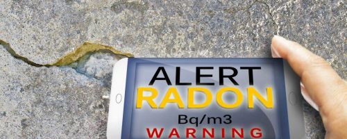
What is Radon?
Radon is an invisible, odourless, naturally occurring radioactive gas. It is a product of the radioactive breakdown (decay) of uranium, commonly found in soil. Radon can enter your home or office through cracks in the foundation walls and floor of your basement, through openings around drain pipes or any other unsealed opening near soil and rocks around your building. When you breathe in radon gas, it gets deposited in your lungs, where it emits radiation. Long-term exposure to high concentrations of radon may lead to lung cancer – particularly if someone in your home is a smoker (http://www.epa.gov/radon/healthrisks.html). In fact, radon exposure is the second leading cause of lung cancer after smoking.
What can I do about Radon in my home or office?
Though most homes do not have a radon problem, the Radiation Safety Institute, Health Canada, the US Environmental Protection Agency and most European countries recommend testing your home for the presence of radon gas, especially if you have a basement. Although you can’t see or smell radon, it can be readily detected with a simple test. If high levels of radon are detected in your house, there are steps you can take to reduce it.
The Radiation Safety Institute and Health Canada recommend conducting a long-term test for radon. This test should last 90 days and should ideally be conducted during the heating season (between October and April, depending on the seasonal conditions in your region of Canada).
Visit www.radiationsafety.ca/community/home-radon-testing for more information.
How is radiation used in medicine?
In medicine, radiation is used both as a diagnostic tool and for treatment (therapy). In diagnostic imaging, X-rays can be used to examine the inside of someone’s body, in order to find, for example, a tumour, a broken bone, a swollen gland, or a swallowed penny. Radioactive materials can be used to produce an image the metabolic activity inside a patient, or the flow of blood through veins, to detect clots or tears (see “What are PET scans?”). Radioactive materials or large quantities of X-rays can also be used in the treatment of cancers, often in conjunction with chemotherapy or surgery.
How do CT scans and Medical X-rays work?
X-rays are high energy electromagnetic radiation. In other words, they are made of the same “stuff” as radiowaves, microwaves, infrared light, visible light, and UV rays, but are higher in energy. Because of their high energy, some X-rays can go right through the less dense parts of our bodies, such as skin and muscle, untouched, like visible light through a pane of glass. These X-rays then reach a film, or detector, placed behind the patient and darken the image. However, the more dense parts of our body, our bones for example, absorb X-rays, preventing them from reaching the film or detector.
As we can see in the X-ray image to the right, all of the x-rays which traveled through the air around the hand reached the film and darkened it. However, very few of the x-rays which had to travel through the thick layer of bone in the wrist reached the film, leaving a very light region in the image. There is a clear contrast between bone and muscle tissue. The muscle tissue is less dense than bone, and so appears darker on the image.
Computed Tomography (CT), also referred to as Computed Axial Tomography (CAT), produces a three-dimensional (3D) X-ray image. Through a series of 2D X-ray images, taken along a ring perpendicular to the subject’s body, a 3D image of the inside of the subject is reconstructed using a computer. CT scans are very helpful to doctors, as they give a complete picture of what is going on inside the body in three dimensions. This allows a doctor to find internal bleeding, or assess exactly where in the body a tumour is while also getting a detailed picture of the size and shape of it. Because CT scans require a series of X-ray images to produce a complete picture, they emit much more ionizing radiation than a typical X-ray does. A CT scan of the chest can give 100 times more radiation dose than a typical X-ray of the same region. However, this radiation dose still results in minimal risk to a patient.
What are PET scans?
Positron Emission Tomography (PET) is a medical imaging tool which uses radioactive substances to produce a 3D image of processes inside a patient’s body. The radioactive substance can either be injected in or ingested by the patient. As it travels through the body, it decays, emitting particles which are captured by the detector. If the substance, for example, is injected into the blood stream, it allows a doctor to track the flow of blood to identify where blood clots are occurring.
PET scans are typically done in addition to CT scans or MRI’s. The PET scan images a process in the body, while the CT scan or MRI gives an anatomical image.
Tell me more about PET scans!
The radioactive substance used in PET imaging is mixed in with another substance, for example a sugar, which travels well through your body. As the radioactive substance decays, it indirectly emits photons which are detected by the ring of detectors. The detectors trace back where the radioactive decay occurred, to build a 3D image of the location of the substance through your body. This imaging modality can, for example, show you how blood is flowing through certain veins, giving a doctor the opportunity to locate clots.
Specifically, the radioactive substance used emits positrons. Positrons are the electron’s anti-particle, which means that when a positron interacts with an electron, the two particles destroy each other (annihilate) and the reaction liberates two photons with specific energies, traveling in opposite directions. Positrons typically do not travel far, after being emitted from a radioactive decay, before annihilating with an electron. The two photons are the radiation captured by the PET detectors. When the detectors record two photons of the right energies hitting different sides of the detector within a short period of time, it is determined that a positron-electron annihilation event occurred, and the position of the event is calculated. It is thereby possible to trace the path of the radioactive material through the body.
How do nuclear power plants produce radiation?
Nuclear power plants use the energy released when heavy particles break apart, or fission, to heat water. The steam produced is sent through steam turbines, creating electricity.
When a uranium atom decays, or splits, it releases high energy photons, a few neutrons, and two fission fragments, the new elements created by the two pieces of what used to be uranium. Radiation is emitted instantly during the fission, in the form of the photons and neutrons, but in most cases, the fission fragments are also radioactive and typically emit both beta and gamma radiation. They could decay almost instantly, adding to the immediate radiation contained in the power plant, or they can remain in their unstable state for long periods of time, only emitting their radiation once they have been removed from the reactor. These materials are the source of high level radioactive waste, and must be segregated and shielded until they are no longer radioactive, which in some cases can take hundreds of years.
The uranium, plutonium and thorium used as fuel in nuclear power plants are not very radioactive by themselves. It is only when large quantities of fuel are surrounded by materials which encourage fission (i.e. in a nuclear reactor) that they become highly radioactive. In this case, a neutron emitted from one fission reaction can hit another heavy atom and break it apart. This induced fission will then produce more neutrons, which themselves can split other fuel atoms, producing a self sustaining chain reaction.
How is radiation used in industry and research?
There are many uses for radiation in industry, from sterilization to testing materials non-destructively. X-rays are used to inspect welds and impurities in materials, and are used for security purposes, for example at airports. Radioactive substances can be used to determine the density and moisture of soil and concrete, for road construction, or to measure flow rates through piping, to list a few examples.
Finally, radiation sources are used to test new materials and probe into the structure of complex molecules. Researchers also try to discover new ways to use radiation in a safe and useful manner, for example to fight disease.
Who regulates the use of ionizing radiation and radioactive materials in Canada?
There are various bodies that regulate the use of radiation in Canada.
The Canadian Nuclear Safety Commission regulates all matters related to radioactive substances and very high energy X-rays. Anyone wishing to purchase, use, store, transport, repair, sell, or dispose of a nuclear substance or radiation device must apply for a licence with the CNSC.
X-ray equipment is regulated provincially. Every province is responsible for setting their own limits and standards. To find the regulations for your province, visit radiationsafety.ca/resources/regulatory-documents.
Health Canada’s Radiation Protection Bureau (RPB) enforces standards for the design and sale of X-ray machines, through the Radiation Emitting Devices Act and associated regulations. Furthermore, Health Canada publishes Safety Codes which outline safety standards for the use of various types of x-ray equipment. These Safety Codes are often used as a basis for provincial regulations.
What is the ICRP?
The ICRP is the International Commission on Radiological Protection. It is an independent, international body that reviews current literature and research on radiation and provides standards and guidelines for radiation protection to the international community. Their guidelines are widely accepted and most countries, including Canada, use them to set national standards, such as yearly limits on radiation dose.
What is EMF?
EMF stands for Electromagnetic Field. The term EMF is commonly misused to refer strictly to very low frequency fields, like the ones which surround power lines and electrical devices. In this case, we are therefore talking about non-ionizing radiation, rather than ionizing radiation like X-rays and the radiation emitted from radioactive particles.
There has been a lot of media attention lately on the question of whether these low energy fields can lead to adverse health effects in humans. This type of radiation has much less energy than visible light and heat, which we know do not pose a health concern. We are therefore not expecting to see the same type of effects from these kinds of radiation than from X-rays and radioactive materials, which we know have enough energy to break electrically neutral atoms into two charged components.
Are cell phones and power lines dangerous?
Cell phones use low-energy electromagnetic fields (microwaves) to communicate. This type of radiation is non-ionizing. Tens of thousands of studies have been done to determine if there are any adverse health effects caused by exposure to EMF. Though most research indicates that there is no link between the radiation caused by low frequency EMF (cell phones and power lines) and health issues, such as cancer, it is still very much an active area of research.
For more information on the current stand of the World Health Organization, Health Canada, and the Health Physics Society, visit www.radiationsafety.ca/community/issues/information-on-emf .
How harmful is radiation exposure?
The dangers of exposure to ionizing radiation depend entirely on the type, energy and amount of radiation you are exposed to.
For chronic exposure to low levels of radiation, the main adverse health effect is an increased risk in developing cancer. The International Commission on Radiological Protection (ICRP) estimates that a radiation dose of 1,000 mSv, received little by little over a long period of time increases one’s chance of developing a fatal cancer by 4%. This estimate is used in setting national occupational dose limits. The Canadian Cancer Society estimates that 25% of Canadians will die of cancer, from all causes, so being exposed to 1,000 mSv of radiation over your lifetime theoretically increases your chance of getting cancer from 25% to 29%.
Sudden exposures to large amounts of radiation bring more immediate effects. Furthermore, these effects are not probabilistic, like cancer, but definite, above a certain exposure. A radiation exposure between 200 and 2,000 mSv leads to a decrease in red and white blood cells and platelets, and will lead to effects such as malaise, vomiting, and diarrhoea. The severity of the effect is proportional to the radiation dose. Half of the people exposed to a dose of 3500 mSv are expected to die if untreated.
How much radiation do I get from a CT scan, and is that dangerous?
As we mentioned in a previous answer, CT-scans are essentially a large set of X-rays, taken at many angles around the body, in order to create a 3-D anatomical image. To put the dose received by a CT-scan into context, remember that on average, Canadians receive 2-4 mSv of radiation dose per year from natural background radiation, which has not been shown to lead to adverse health effects, and that an accumulated exposure of 1000 mSv over a lifetime leads to an estimated 4% increased risk of developing cancer.
We know a single X-ray does not deliver much radiation dose: a typical chest X-ray delivers about 0.1 mSv of radiation, about the same radiation dose you receive from background radiation in about 12 days. However, CT-scans are a series of many X-rays, and deliver much more radiation dose than a regular x-ray. A typical chest CT, for example, delivers 6 mSv of radiation dose. Though this may triple your annual radiation dose for that year, this still represents a very small risk You would need about 40 CT-scans in your life to increase your chance of getting a fatal cancer by 1%.
Scans over larger areas, like the pelvis, abdomen and chest, can give about 10 mSv of dose, whereas a typical head CT may only give 1.5 mSv. A screening mammogram delivers about 3 mSv of radiation dose.
Still, as it is assumed for safety reasons that any level of radiation dose leads to an increased risk of developing cancer, doses must be kept as low as reasonably achievable, and CT scans should only be administered if the potential benefits from getting a scan (e.g. finding a suspected tumour) far outweigh the possible adverse health effects from the exposure to radiation. In other words, doctors need to have justification for ordering a scan.
How much radiation do I get at the dentist, and is that dangerous?
It may make you uneasy to have an X-ray machine pointed at your face every time your dentist wants an updated image of your teeth. But in reality, especially with today’s technology, the radiation dose received from dental X-rays is very, very small, and the possible benefit from the X-ray image (e.g. finding a cavity, locating wisdom teeth to determine if they are ready to be pulled, etc) far outweigh the possible adverse health effects from the exposure.
The Health Physics Society, a large body of radiation protection professionals in the United States, ascertains that “for a dental panoramic radiograph, the effective dose is [0.026 mSv], which is the equivalent of about 3.3 days of natural background radiation. A series of four [dental X-rays] is [0.038 mSv], which is the equivalent of 4.8 days of background radiation.”
When you are in a plane, you are exposed to more cosmic radiation (radiation emitted by the Sun) than you are when you are on the ground, since you are higher up in the atmosphere. It is calculated that a typical cross country flight delivers about 0.030 mSv of additional radiation dose to passengers, so a series of dental x-rays gives about the same radiation dose as one cross-country flight.
Can I become radioactive if I’m around radiation?
The answer to this question is yes and no. It depends on the type of radiation that you are exposed to and what type of exposure you receive. As we saw in “What is radiation?”, there are two types of ionizing radiation (radiation which is energetic enough to break atoms apart): radiation emitted from radioactive materials, which can come in the form of particles or photons (bundles of energy), and man-made radiation, such as X-rays, which are also photons.
Let’s start with the easy one: X-rays. X-rays cannot make materials, or humans, radioactive. X-rays are very much like visible light from a light bulb. When the light bulb is off, no light is being emitted. Similarly, if an X-ray unit is off, or even on stand-by, no X-rays are being emitted. Furthermore, most materials, and certainly humans, do not emit their own light when they are exposed to visible light. It is again the same with X-rays; when X-rays traverse our bodies, they are either break bonds in the molecules in our bodies, in which case they immediately dissipate their energy and the damage is instantaneously done, or they go right through us, and nothing happens to our bodies. As soon as the X-rays have interacted or passed, the radiation is gone. Nothing becomes radioactive because of X-rays. If the X-ray producing unit is off, there is instantaneously no more radiation around, anywhere.
However, if radioactive materials are ingested, inhaled or absorbed by your body, then you do become radioactive, in a way. As the radioactive material decays in your body, which can occur quickly or very slowly, depending on the material, it will emit radiation which will either deposit dose in your body or, if the radiation is in the form of a gamma ray, it will exit your body, potentially giving dose to a nearby person. This is why patients that are treated with radioactive materials, for example for thyroid cancer, are told to avoid hugging others, or sharing a bed for a few days, until the radioactive material has either mostly decayed or exited the body.
Are the new full-body scanners used at airports safe?
A short answer to this question is that full body scanners are safe. The controversy with full-body scanners is not related to the radiation dose, but with the revealing images that they produce.
There are two types of Full Body Scanners: Millimeter Wave Scanners, used in Canada and which give absolutely no radiation dose, and Backscatter X-ray Machines, used in the USA and elsewhere, which use very low energy X-rays to produce an image the surface of the body. The latter does give a small radiation dose, but this dose is less than that received during a dental X-ray. It is also less than the dose the passenger will receive on a typical flight. Indeed, at higher altitudes, the radiation dose from cosmic radiation (high energy radiation from the Sun) is increased, giving about 0.005 mSv of dose for every hour on board. The dose received from a Backscatter X-ray is on the order of 0.001 mSv.
On the right is a picture of a Millimeter Wave Scanner, which produces a 3-D image of the surface of your body in less than a few seconds. A Backscatter X-Ray machine looks more like a wall which you stand in front of, and produces a 2-D image of the surface which faces the wall.
For a complete answer to this question, visit www.radiationsafety.ca/airport-backscatter-x-ray. Since this subject has received a lot of press in recent years, we’ve dedicated a full webpage to it!
Don’t see the topic you are wondering about? Let us know, and we will personally answer your questions.